At the UI Central Microscopy Research Facilities, their powerful microscopes are like the Hubble in reverse: dynamic scientific tools capable of drawing out beauty on an extremely small scale.
To celebrate the artistry found in nanometers, the Iowa Microscopy Society will hold its annual “Iowa Art in Science” competition on Wednesday, Sept. 28. The competition is held in conjunction with the Iowa Microscopy Society Symposium, hosted this year at the University of Iowa. The deadline for submission is Sept. 21. Contest entry form and additional information about the Iowa Art in Science competition can be found here.
“I think we all as staff have a bit of an artist eye,” said Tom Moninger, assistant director of the UI Central Microscopy Research Facility. “Images will be more effective in a journal if it catches the eye and tells the story of your research. Plus I have some investigators that love it when I see something really weird and just take a picture of it. Suddenly that gets the gears going – what is going on there? – and then leads to other discoveries.”
Symposium attendees will vote on the winners. Winning submissions will be featured in the 2017 UI Central Microscopy Research Facility calendar and will receive prizes donated by corporate supporters of the Iowa Microscopy Society Symposium.
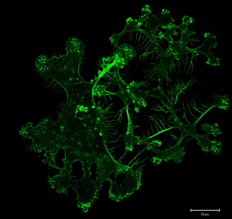
Associate Research Scientist Karla Daniels, UI Department of Biology, is a past winner of the Iowa Art in Science competition whose work has also won photomicrography competitions at Nikon and Bio-Rad. She said that considering the artistic side of the scientific work she pursues has made her appreciate her research in new ways.

The UI Central Microscopy Research Facility is one of the leading university microscopy facilities in the nation supporting outstanding scientific discovery among biological and physical science investigators. It is part of the Office of the Vice President for Research and Economic Development, which provides resources and support to researchers and scholars at the University of Iowa and to businesses across Iowa with the goal of forging new frontiers of discovery and innovation and promoting a culture of creativity that benefits the campus, the state, and the world. More at http://research.uiowa.edu, and on Twitter: @DaretoDiscover.
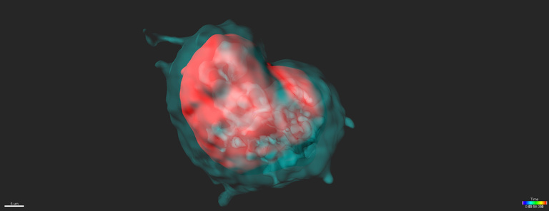
Photos (from top): An image of a primary human dendritic cell captured by Karla Daniels from David Soll’s lab, awarded Image of Distinction in the 2007 Nikon Photomicrography Competition; Postdoctoral Associate Lu (Nemo) Lin submitted this heart shaped image of expanded banana slug mucus which was a winner in the 2015 Iowa Art in Science competition. Both images were taken using UI Central Microscopy Research Facility equipment.