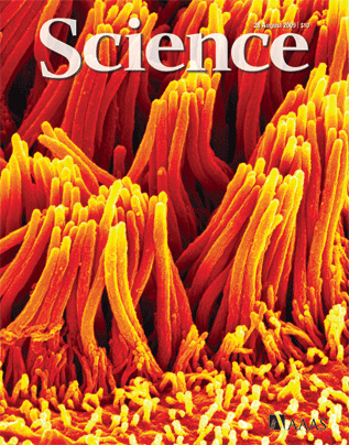
The UI Central Microscopy Research Facility (CMRF) is equipped with two things critical to any researcher’s needs: millions of dollars of top-of-the-line, well-maintained microscopy equipment and a dedicated, knowledgeable staff who specialize in the design, execution and analysis of microscopy experiments.
Six staff members help researchers get the most out of this UI core research facility. Director Randy Nessler and Assistant Director Tom Moninger both got involved with microscopy as student employees in the 1980s, washing glassware used to hold specimens.
“You kids don’t know how easy you have it. When we started working here it was all film–wet chemistry,” Moninger said, laughing. “When we wanted to take a picture of a fluorescently labeled sample, we’d a number of pictures and then send the film down to University Camera. We then had to wait a day or two, and only then did we know if we got good results or not.”
“Three days’ lag and then ‘Oh, it’s out of focus. Do it again,’” said Nessler dryly. “My current cellphone blows away the original top-of-the-line office computers we first installed in the 1980s. Everything is digital now–instant gratification.”
Related: Grant applications open for Central Microscopy Research Facility seed funding
The small CMRF staff is big on experience. Several staff members have worked in the CMRF for more than 20 years and are top in their field. While every member of the CMRF staff is generally knowledgeable about every piece of equipment, each microscopy microscope in the lab has a staff member who “…knows that machine inside and out,” Moninger said.
Mei-ling Joiner, Assistant Research Professor, Molecular Physiology and Biophysics, has capitalized on that experience. Last fall, she was awarded a grant through the CMRF’s Pilot Project Seed Grant Program to develop a new line of research into Parkinson’s disease. Under a tight deadline to apply for another grant using preliminary data from this experiment, Joiner said CMRF Assistant Research Scientist Chantal Allamargot’s expertise was critical.
“The money I got from the seed grant was enough to do one run with the experiment, so it had to work,” Joiner said. “The experiment had to be precise and well-constructed. Working with the people in the CMRF made it possible to get it to work on the first time through.”
The seed grant funding paid the lab fees for using the equipment and for “consumables” used to conduct the experiments, including special papers, stains and dyes. It also covered hands-on training that Allamargot gave Joiner in new techniques that worked well for her research goals. Allamargot even showed her how to locate certain landmarks in Joiner’s specimens that would point to the neurons she was trying to isolate.
“The new techniques blended with some old techniques I had learned previously enabled me to get this data done in about a month, which is pretty fast for getting useful data,” said Joiner, who is now continuing this line of research with a grant through the Carver College of Medicine. “To do the same work on my own without assistance from Chantal, the learning curve would have been a lot longer. I probably would have missed my grant deadline and then I’d have to wait another year. By then it could have been too late.”
While some university-based core microscopy facilities focus on one particular kind of microscopy–such as confocal or electron–the CMRF is equipped to support the majority of requests from local researchers whether they are biological investigators or physical science researchers. The facility includes a vast array of microscopy equipment: transmission electron, scanning electron, confocal/super resolution, and sample preparation. It also has a bioluminescence imaging machine that can image a whole living animal, allowing investigators to document the growth or regression of tumors over time.
Additionally, thanks to a generous contribution from the Roy J. Carver Charitable Trust, two years ago the CMRF added a Stimulated Emission Depletion (STED) Super Resolution Microscope to its arsenal of equipment. The STED system can resolve structures five times smaller than the theoretical limit of a light microscope. It’s the only one of its kind in the state of Iowa. Researchers would have to travel to Kansas City or Madison, Wis. to access the next closest one.
Moninger encourages investigators to meet with CMRF staff as they prepare external grants to understand the costs and potential implications of their projects.
“Then the investigator can demonstrate to the granting agency that we have the tools available to get the work done plus the preliminary data,” he said, adding that CMRF staff want to help investigators advance their research through collaboration on experiment design, preliminary data gathering, publication assistance, grant application support letters, and even journal cover art.
“When you are writing a grant, the more support letters to show that you can do the experiments the better,” said Joiner. “The Iowa Central Microscopy facility has an outstanding reputation and if you get a letter from the director saying you have the ability to do the experiment and experiments like these are just routine for them, that is a big deal for support on your grant.”
The UI Central Microscopy Research Facility is one of the leading university microscopy facilities in the nation supporting outstanding scientific discovery among biological and physical science investigators. It is part of the Office of the Vice President for Research and Economic Development provides resources and support to researchers and scholars at the University of Iowa and to businesses across Iowa with the goal of forging new frontiers of discovery and innovation and promoting a culture of creativity that benefits the campus, the state, and the world. More at http://research.uiowa.edu, and on Twitter: @DaretoDiscover.
Photo: This 2009 Science magazine cover image highlights the cilia that line human trachea. It was acquired on a Scanning Electron Microscope at the UI CMRF by Tom Moninger for the Michael Welsh lab.